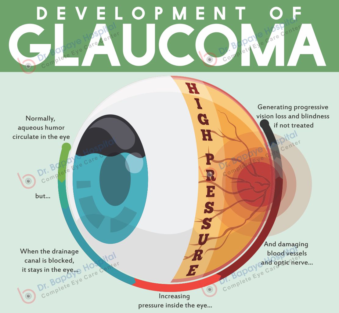
Glaucoma is often known as a thief of vision as a patient may lose vision gradually even without having a single symptom. Vision lost due to glaucoma is permanent and cannot be regained. Early diagnosis and follow-ups at regular intervals are the only way to preserve the vision and avoid further vision loss. The intraocular pressure (IOP) is raised causing damage to the optic nerve. Sometimes optic nerve damage can occur even with seemingly normal IOP. The vision loss is irreversible and starts from the periphery and so is not noticed early. Hence all patients are screened for glaucoma. Patients suffering from glaucoma are regularly tested using various instruments and parameters.
At Dr. Bapaye Hospital, we are the best Glaucoma Clinic in Nashik offering state of the art diagnostic and therapeutic facilities for glaucoma patients.
Diagnostic Facilities for Glaucoma
Applanation Tonometer (IOP)
The gold standard for measurement of intraocular pressure (IOP). This is done on every visit for every patient above 40 years.
Have questions about glaucoma clinic?
Talk to our specialists — ethical advice, no obligation.
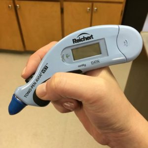
Tonopen
A handy device for IOP measurement in a special group of patients like children and physically invalid.
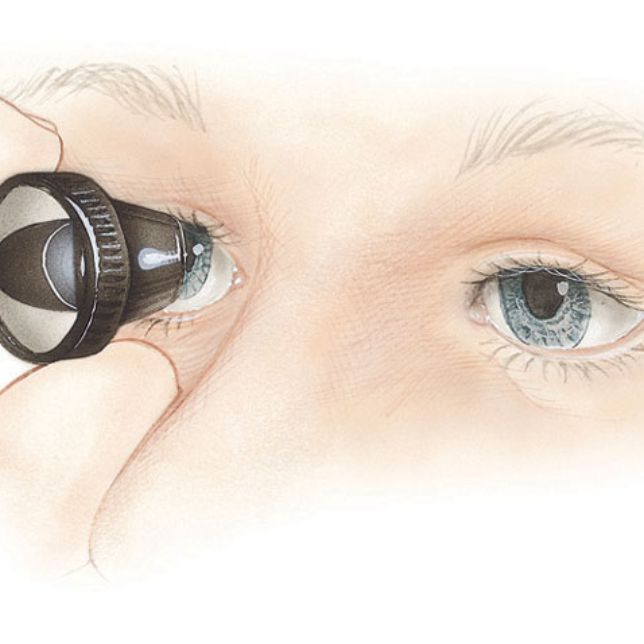
Keeler Non-Contact Tonometer
An accurate method to measure IOP in the busy outpatient department to screen each and every patient for glaucoma.
Gonioscopy
For accurate assessment of anterior chamber angles. This test is done to differentiate between the two types of glaucoma i.e. Open-angle and Closed-angle type.
Open-angle Glaucoma is managed first by medication, and if uncontrolled then surgery (Trabeculectomy). Closed-angle glaucoma is medically controlled, then a laser treatment called Peripheral Iridotomy (Yag PI)is done and if required surgery is done.
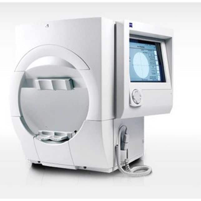
Humphrey Field Analyzer (Zeiss)
Automated perimeter for assessment of visual fields.
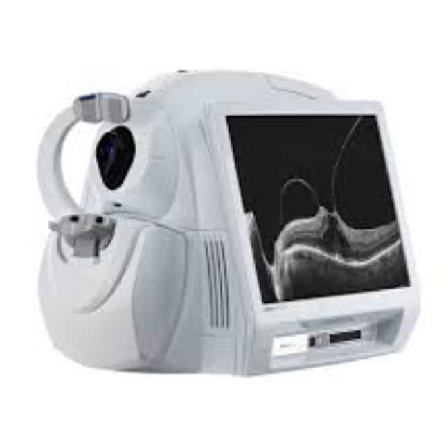
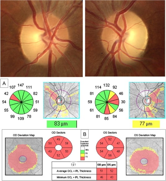
Cirrus- HD OCT (Zeiss)
The latest software is capable of anterior and posterior segment assessment. Anterior segment features include pachymetry and outflow angles measurement while the posterior segment features do an assessment of the optic nerve and RNFL assessment.
Forum Workstation
For comprehensive glaucoma analysis by using HFA and OCT data. Immensely useful for serial assessment and prediction of glaucoma progression.
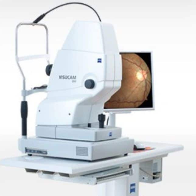
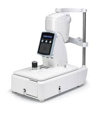
Zeiss FF 450 IR Fundus Camera
For photography of optic nerve head and retinal nerve fiber layer
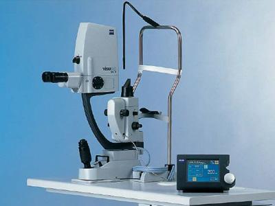
YAG Laser
To perform peripheral iridotomy in narrow-angle glaucoma.
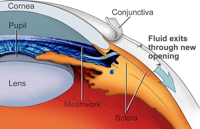
Trabeculectomy and Glaucoma Implant Surgery
For advanced glaucomatous eye disease.
Iridex Red diode laser (GLX 810) with G-probes
For cyclophotocoagulation in advanced glaucoma. This procedure is required for symptomatic relief in painful blind eyes.
OUR SERVICES
What causes glaucoma?
Glaucoma is usually caused by raised pressure inside the eye (intraocular pressure) damaging the optic nerve, though it can occur even with normal pressure. Age above 40, family history, diabetes and long-term steroid use increase risk. Because early glaucoma has no symptoms, every patient at Bapaye Eye Hospital is screened for it during a routine eye check-up.
Can vision lost to glaucoma be restored?
No — vision lost to glaucoma is permanent, which is why it is called the silent thief of sight. Treatment with drops, laser or surgery can only preserve the vision you still have. That makes early detection and regular follow-up examinations the single most important step for anyone over 40 in Nashik.
How is glaucoma treated at Bapaye Eye Hospital?
Treatment depends on the type and stage: pressure-lowering eye drops first, laser procedures (iridotomy or trabeculoplasty) where indicated, and surgery such as trabeculectomy or glaucoma implants for advanced cases. Our glaucoma clinic includes a fellowship-trained consultant from Sankara Nethralaya, Chennai.