Vitreoretinal surgery refers to superspecialty of ophthalmology involved in treatment of eye diseases involving the retina, macula, and vitreous fluid. Retina is the sensitive layer situated at the back of the eye. The light coming from an external object is focused by the cornea and the lens onto the retina. Within the retinal cells the light is converted to electric impulses/neural signals which are then taken to the brain via optic nerve.
Diseases that affect the retina are often a reflection of the general health of an individual. Retina gets affected by various systemic diseases like diabetes mellitus, hypertension, etc. A large number of diseases affect the retina which requires both medical as well as surgical treatment.
Common Retinal Diseases:
Retinal detachment
Retinal Detachment: Important Things You Need to Know
In our last blog we have discussed the structure of the eye. However just to refresh our memory, an eye works like a camera. The light coming from an object is focused by the cornea and the lens, which act as focusing mechanisms, onto the film of the eye, known as the retina. The space
Read More »Have questions about vitreo-retinal services?
Talk to our specialists — ethical advice, no obligation.
Watch now for Vitrectomy for retinal detachment
Age related macular degeneration
Age-Related Macular Degeneration
Age Related Macular Degeneration Dr.Maneesh Bapaye DNB, FRCS(G), FMRF,FICO,MNAMS,MBA Fellow-Sankara Nethralaya, Chennai Consultant Vitreoretinal Surgeon, Dr.Bapaye Hospital, Nashik Age-related macular degeneration (AM.D.) is a major cause of blindness in patients above the age of 55-60 years. The disease is more prevalent in western countries but is on the rise in the Indian subcontinent as well.
Read More »Diabetic Retinopathy
Diabetic Eye Disease & Diabetic Retinopathy
Diabetes mellitus is a lifestyle disease that is rapidly increasing all over the world. India has the dubious distinction of being the diabetes capital of the world. Diabetes causes damage to all the organs in the body. Eye also gets affected in diabetic patients. Patients with diabetes get Lets us understand more about diabetic retinopathy
Read More »Watch now for Vitrectomy for advanced diabetic retinopathy
Watch now for Ozurdex Injection for Macular Oedema
Macular hole
What is a macular hole?
A macular hole is a condition where a circular opening or break forms in the macula. The macula is the central part of the retina and is responsible for sharp, central vision, which is necessary for tasks like reading and driving. How is macular hole formed? The primary cause of a macular hole is age-related
Read More »Watch now for Vitrectomy for Macular Hole
Epiretinal Membrane
What is Epiretinal membrane?
An epiretinal membrane (ERM) is a thin layer of tissue that forms on the surface of the retina, the light-sensitive layer of tissue at the back of the eye. The membrane is composed of cells and fibers and can cause distortion or blurriness of vision. Epiretinal membrane can develop as a result of age-related changes
Read More »Watch now for VITRECTOMY FOR ERM REMOVAL
Retinopathy of Prematurity
Retinopathy of Prematurity
Dr. Maneesh Bapaye Dr. Bapaye Hospital, Nashik Retinopathy of prematurity (ROP) is an eye disorder that affects premature infants. It occurs when the blood vessels that supply the retina, the part of the eye that detects light, grow abnormally. ROP can cause vision loss and even blindness if left untreated. There are several risk factors
Read More »Floaters
What are floaters and how are they treated?
Small, moving specks called eye floaters can occur in your range of vision. They can be solid, semi-solid, transparent, or take on other shapes like dots, circles, lines, or cobwebs. They appear to be floating in your eye and move in response to eye movement. Changes in the vitreous, a material that resembles gel and
Read More »Watch now for Vitrectomy for floaters
A wide variety of diagnostic devices as well as treatment equipment is essential to achieve optimum results in retinal diseases.Dr. Bapaye Hospital has a state of the art Vitreo-retina department well equipped with all the diagnostic and therapeutic instrumentation and expertise necessary to manage any retinal problems.
Diagnostic equipments for Retinal diagnostics
Zeiss Fundus Camera
Zeiss Fundus Camera- Color Photos, Autofluorescence, Red Free Photography Flurorescein angiography and ICG angiography of the retina
OCT Cirrus 6000
Zeiss Cirrus 6000 OCT and OCTA- Optical Coherence Tomography (OCT) is a non-invasive imaging test that uses light waves to take detailed cross-sectional pictures of the retina. OCT Angiography (OCTA) is an advanced version that maps blood flow in the retina, helping doctors detect and monitor eye conditions like diabetic retinopathy or age-related macular degeneration without the need for dye injections
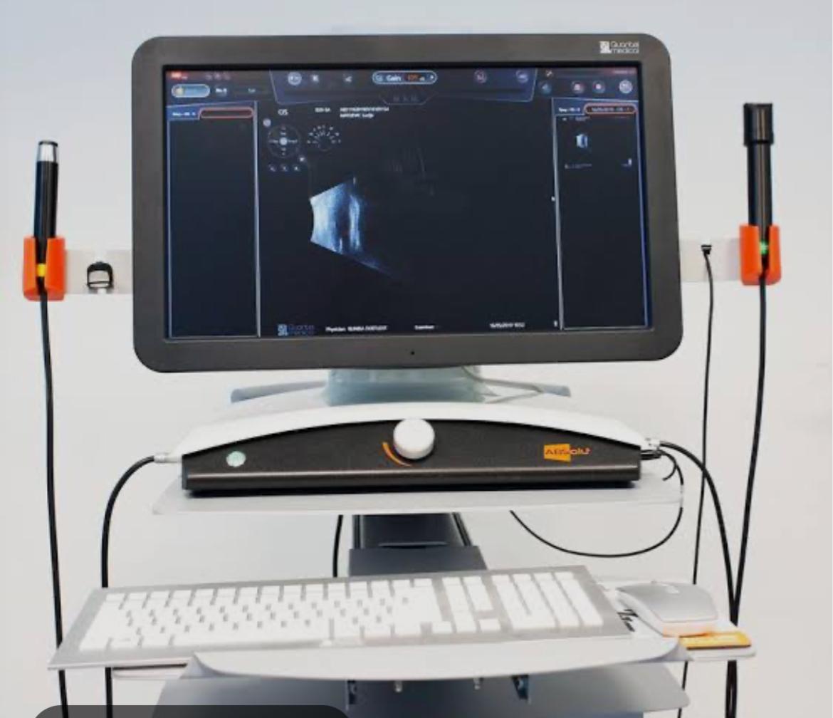
Ophthalmic Ultrasonography
Ophthalmic ultrasonography ( Quantel medical)- this eye test that uses sound waves to create detailed images of the inside of your eye. It is performed when the retina can not be visualized like in cases of dense vitreous hemorrhage or mature cataract. It can also be performed to assess structures in the back of the eye as in cases of retinal detachment or suspected cancer of retina or choroid. The test is painless and usually takes only a few minutes
Surgical Services for Vitreoretinal disorders
Constellation Vision System (Alcon Inc)
Constellation vision system allows the surgeon to perform high-speed, transconjunctival sutureless vitrectomy to achieve accurate surgical results with minimal surgical time. All types of retinal surgical procedures like macular surgeries, retinal detachment, diabetic vitrectomy, retinopathy of prematurity, etc
Zeiss Green Laser System
Zeiss Green Laser system is used for retinal lasers in outpatient clinic
Iridex Green Laser System
Iridex Green Laser system is used for performing retinal laser during course of retinal surgery
Zeiss Resight Visualisation System
Zeiss Green Laser system is used for retinal lasers in outpatient clinic
Intravitreal Injections of Anti VEGF drugs such as Eylea, Pagenex, Accentrix, Razumab and Avastin etc are available for treatment of diseases like Age Related Macular Degeneration (AMD), Choroidal Neovascular Membrane (CNVM)
Diabetic retinopathy and diabetic macular edema (DME), Vitreous hemorrhage, Retinal Vein Occlusions, Retinopathy of Prematurity (ROP), Neovascular Glaucoma etc
Intravitreal steroid injections like Ozurdex are used for diabetic macular edema, Cystoid Macular Edema (CME), Uveitis
Watch now for Retinal Angiography
Watch now for Laser procedure for Retinal diseases
Watch now for Optical Coherence Tomography (OCT) non invasive investigation for retinal diagnosis
Looking for VITREO-RETINAL SERVICES?
Fill your details here. You will receive a call from our patient care desk.
OUR SERVICES
Cataract
LASIK & Refractive Surgery
Vitreo-Retinal
Pediatric Ophthalmology & Squint Services
Glaucoma Care
Cornea Clinic
Contact Lens
Other Services
How often should a diabetic get their retina checked?
At least once a year, even with perfect vision — diabetic retinopathy damages the retina silently before symptoms appear. Patients with longer diabetes duration or known retinopathy need more frequent reviews. Timely laser or injections control the disease; vision already lost to advanced retinopathy often cannot be recovered.
What are the warning signs of retinal detachment?
Sudden floaters, flashes of light, or a curtain-like shadow across your vision are emergencies — the retina may be detaching. Call 0253 2506505 immediately; surgery within days protects central vision. Dr. Maneesh Bapaye (Fellow, Sankara Nethralaya) performs vitrectomy and retinal detachment repair at our centre.
Are eye injections for retina safe?
Intravitreal injections (anti-VEGF) are a standard, well-tolerated treatment for diabetic macular edema and vein occlusion, done under sterile operation-theatre conditions in minutes. Most patients need a series of injections; your retina specialist plans the schedule based on scans.