
CORNEA CLINIC IN NASHIK
The cornea is the transparent front portion of the eye that covers the iris, pupil, and anterior chamber. The cornea, with the anterior chamber and lens, refracts light, with the cornea representing roughly 66% of the eye’s total optical power. Corneal blindness is a very common cause of avoidable blindness in our country.
Dr. Bapaye Hospital serves as a referral center for management of complex corneal problems like infective corneal ulcers, chemical burns, unresponsive allergic conjunctivitis from the city as well as neighboring villages.
Dr. Bapaye Hospital offers Corneal topography and C3R for keratoconus, as well as lamellar and penetrating keratoplasty (corneal transplants) for corneal diseases.
We are Best Cornea Clinic in Nashik with a well-equipped cornea eye bank that is being used for treating patients with corneal disorders.
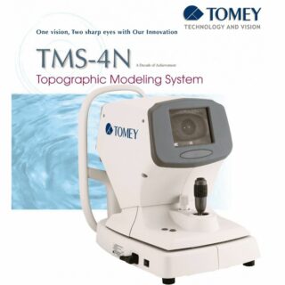
Corneal Topography system used for measurements of corneal curvature and aberrations. It is employed in patients suspected of having corneal abnormalities like Keratoconus.
Corneal topography provides more data, quantitates corneal shape, and measures patterns of irregular and induced astigmatism. Corneal topography used to be considered a specialized test to diagnose corneal pathology or to help fit contact lenses, but since the introduction of laser vision correction in the mid-1990s, it has become a common test used to screen potential surgical candidates. With the refinements in cataract surgery techniques and technology, particularly advanced IOLs, corneal topography is an essential part of the preoperative workup. In addition, it is valuable for detecting subtle irregularities of the corneal epithelium (i.e., anterior basement membrane dystrophy) and tear film (dry eye).
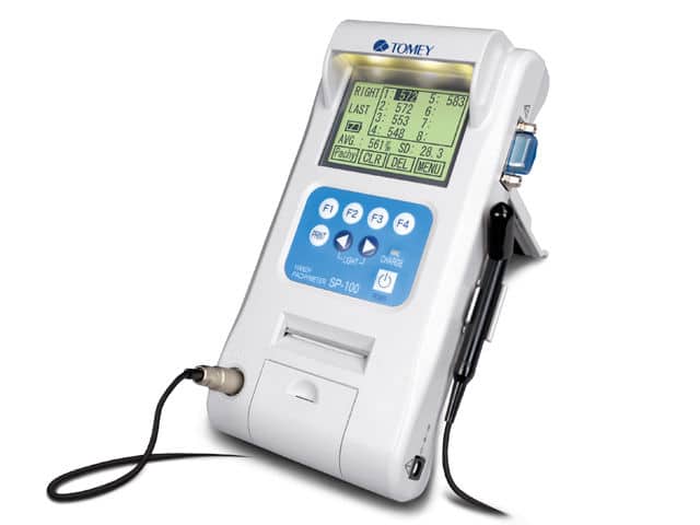
For measurement of corneal thickness.
Corneal Collagen Cross-Linker (Eyeroc UVX-1000)
The device was developed by the pioneer of corneal collagen cross-linking, Prof. Theo Seiler. This is used in the treatment of keratoconus to stop the progression of disease and stabilizing refractive error.

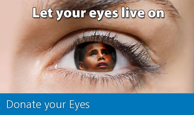
Add Color to One’s Life, Donate Eyes
The cornea is the clear tissue covering the front of the eye. It serves as a window to allow light to enter the eye. If the cornea becomes cloudy from disease, injury, infection, or poor nutrition, vision is dramatically reduced or lost. Corneal blindness affects mainly children and young adults who have a long life ahead of them.
Procedure to Donate Eyes
After death and should be collected within 6 hours. Eyes will be collected at the donor’s place. Collection of eyes will take only 10-15 minutes. A trained physician using a sterile procedure removes the eyes. To ensure no disfigurement of the face, artificial eyes are fitted in place. 10 cc of blood is collected for testing.
Who can Donate Eyes
Practically anybody can donate from the age of 1 year. Even if the deceased has a medical history of hypertension, diabetes, asthma, tuberculosis etc can donate. Spectacle wearers and people who have undergone cataract operations can donate eyes.
Ocular surface
- Pterygium is a fleshy growth on to the cornea which causes development of cylindrical power, decreased vision, redness, watering and irritation. It needs surgical removal. We offer the best type of pterygium surgery with Autograft with imported Fibrin glue, which has the best outcome and least chance of recurrence.

- Chemical injuries like lime and acid burns need emergency care. They also sometimes need ocular surface surgery with amniotic membrane and stem cell transplant.
