
A simple, friendly guide for patients and families — what retinal detachment is, its warning signs, how it is treated, and what to realistically expect from recovery.
🚨 Sudden floaters, flashes or a shadow? Call 0253 – 2506505 todayEver wondered how your eye creates the pictures you see? The simplest way to understand it is to compare your eye to a camera — because they work in almost exactly the same way. Both focus light onto a light-sensitive surface to form an image.
When you look at a tree, light bounces off it and enters your eye through the clear front surface — the cornea (the camera's glass lens). It then passes through a natural lens that fine-tunes the focus, just like a camera's zoom. At the very back sits a thin, delicate lining — the retina — your eye's "film" or digital sensor. The retina catches the focused light and converts it into signals that travel through the optic nerve to your brain. That is what you "see."
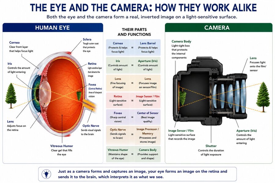
Just as a camera cannot take photos if its sensor is damaged or loose, your eye cannot see properly if the retina is damaged or detached. That is exactly what this guide is about.
Before we talk about what goes wrong, here's a quick, friendly tour of your eye. Don't worry — we'll keep it simple.
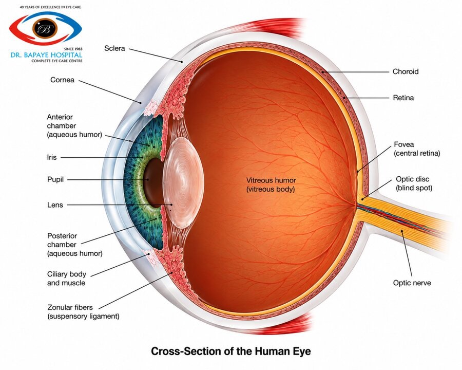
The clear, dome-shaped front window of your eye. It's what light first hits when it enters your eye.
The coloured part (iris) with a central hole (pupil) that widens in dim light and shrinks in bright light — just like a camera aperture.
A flexible, transparent disc just behind the pupil. It fine-tunes the focus of light onto the retina.
A clear, jelly-like substance that fills most of the inside of the eye — like the stuffing inside a cushion — giving the eye its round shape.
The thin, light-sensitive lining at the back — your eye's "camera film." This is the star of our story.
The very centre of the retina, responsible for your sharpest vision — reading, recognising faces, seeing fine detail.
The tough, white outer coat of the eye — the protective shell that holds everything together.
The cable that carries visual signals from the retina to the brain, where they are understood as images.
Imagine taking a beautiful photograph, but the film inside the camera is torn or crumpled. No matter how good the camera is, the photo comes out ruined. The retina is exactly like that film.
Your retina is wafer-thin — thinner than a strand of hair — yet it contains millions of tiny light-sensitive cells called rods and cones that turn light into electrical signals your brain can understand.
Reading, recognising faces, watching TV, threading a needle (the macula).
Sensing movement around you and walking safely (peripheral vision).
Seeing the world in all its colours.
Seeing in low-light conditions.
The retina is attached to a layer beneath it called the choroid, which supplies it with blood and nutrition. As long as the retina stays flat against the choroid, your vision is nourished and works well. When the retina separates from the choroid — that is a retinal detachment — it is cut off from its blood supply and begins to die.
When we are young, the clear jelly (vitreous) is firmly attached all along the inner surface of the retina — like wallpaper glued perfectly to a wall.
As we get older (usually after age 50–60), this jelly naturally shrinks and becomes more watery. As it does, it slowly peels away from the retina — starting from the back and working forward. This natural, age-related process is called Posterior Vitreous Detachment (PVD) — or simply, "the jelly separating from the lining."
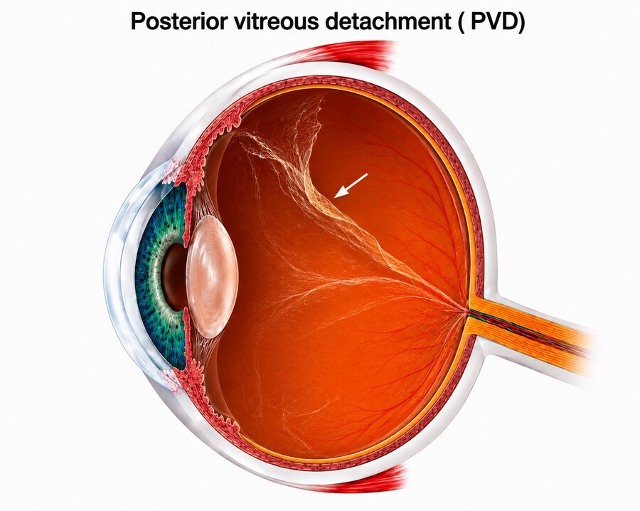
Many people notice these symptoms when PVD happens. They are usually harmless but should be checked by a doctor:
Sudden appearance of black spots (floaters)
Flashes of light, like lightning inside the eye
Cobweb or curtain-like shapes in vision
A large ring-shaped floater
In most people PVD happens smoothly. But in some, the jelly is extra sticky and firmly glued to certain spots. When it tries to pull away, instead of letting go cleanly it tears the retina — like pulling double-glued wallpaper and taking a chunk of wall with it.
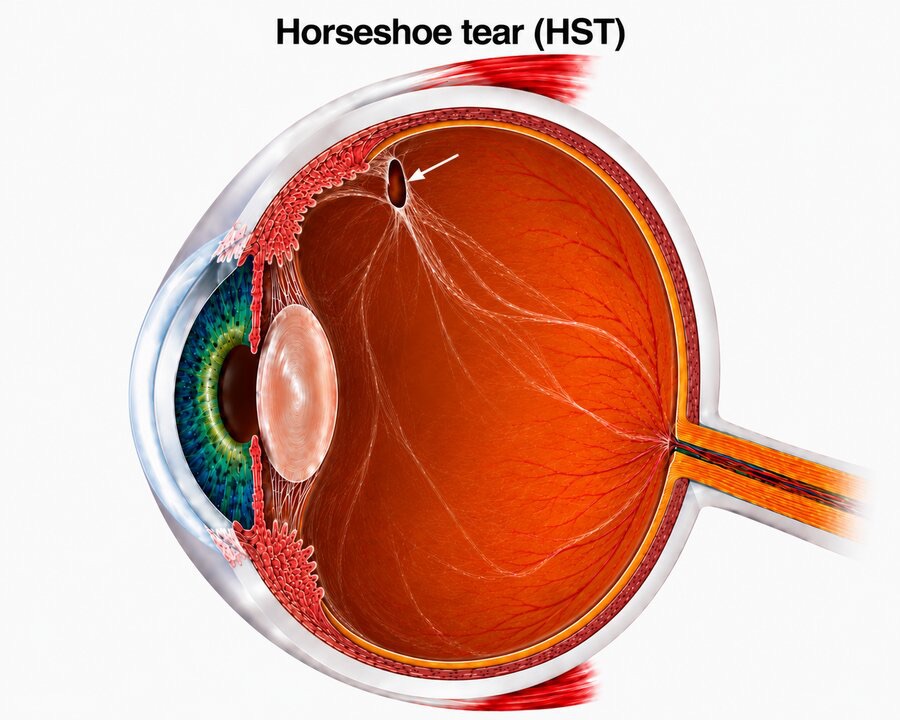
This tear is called a Horseshoe Tear (HST) because of its U-shape. A small flap of retina is still being tugged by the attached jelly. Some people also have areas of naturally thin, weak retina called lattice degeneration — like patches of worn-out wallpaper — which are more prone to tearing.
Once a horseshoe tear or a hole forms, a dangerous chain of events can follow if left untreated. Tap through each step:
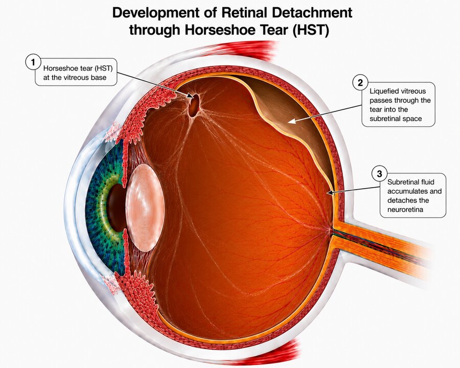
A tear forms in the retina — like a small hole punched in a boat's hull.
Fluid from the vitreous jelly seeps through the tear and gets behind the retina — like water leaking into the hull.
This fluid accumulates and pushes the retina away from its base — like wallpaper bubbling off a wet wall. This is the retinal detachment.
Separated from its blood supply, the retina begins to lose its function. The patient sees a dark shadow or curtain.
If the macula (centre of the retina) detaches, central vision drops suddenly and significantly.
Words can only describe so much. This shows — from a patient's own point of view — how vision changes when retinal detachment occurs, and what it can look like after successful surgery.
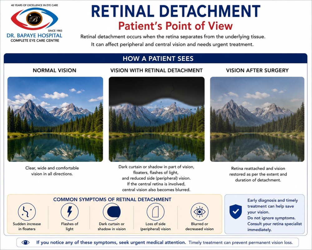
Many patients describe "a dark curtain falling across my vision" or "a shadow from the corner that kept growing." Some also see multiple black spots drifting across their field of view, or bright flashes like lightning — especially in a dark room.
A storm of new black spots or cobwebs appearing suddenly
Like camera flashes or lightning bolts inside the eye
A growing dark area blocking part of your vision
Like wearing blinkers — side vision narrows or disappears
Overall dimness or blurring that appears suddenly
If a retinal tear is found before significant detachment has occurred, your doctor can seal it with a simple, painless laser treatment done right in the clinic — no operation theatre needed.
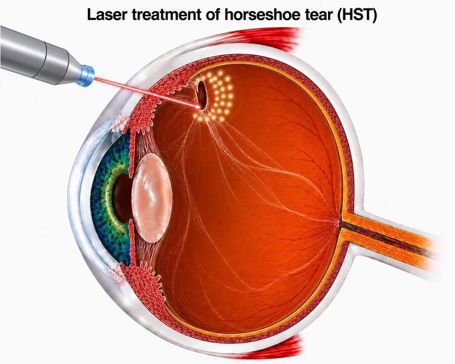
The laser creates tiny, controlled burns in a ring around the tear. These act like spot-welding — they create scar tissue that glues the retina firmly to the underlying layer, so fluid cannot creep behind it.
Prevent a tear from progressing to a full detachment · treat small, early detachments that haven't reached the centre · "wall off" the area in early stages · act as a preventive measure in high-risk patients.
After laser, the tear is sealed but floaters may remain for some time — they are harmless remnants. A very small number of patients may still develop detachment despite laser, especially in the first 2 weeks before the seal is complete. Follow-up as advised is essential.
For select patients, a small gas bubble is injected into the eye to push the retina back into place — no surgery theatre, no big incisions.
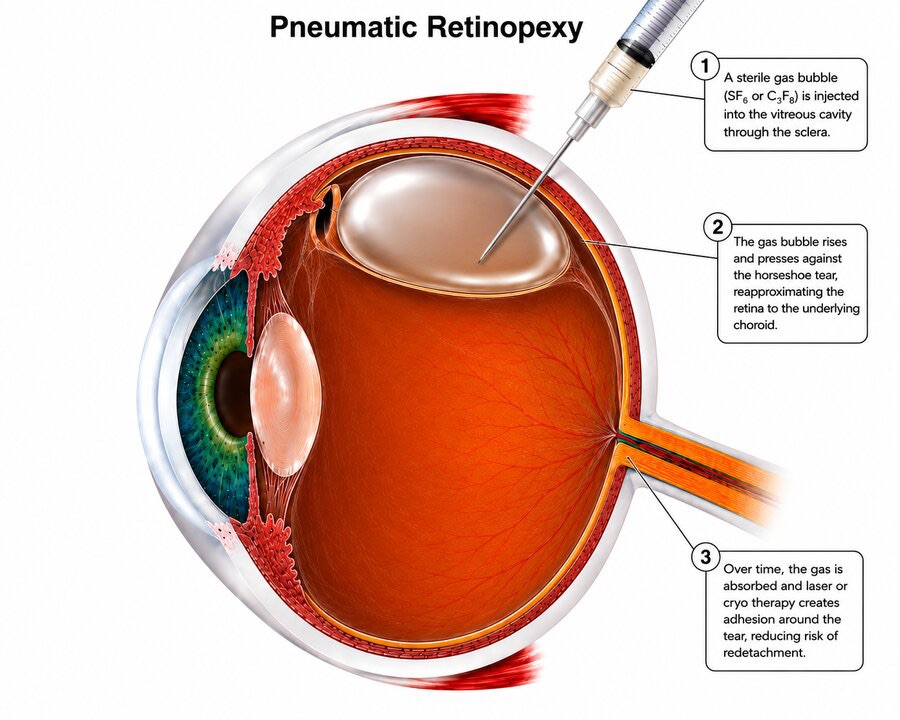
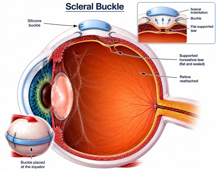
One of the most time-tested and effective surgeries for retinal detachment, used for decades with excellent results. Think of it as gently reshaping the eye from the outside to bring the wall back to the wallpaper.
Vitrectomy is the most commonly performed surgery for retinal detachment today. While the scleral buckle works from the outside, vitrectomy works from inside the eye — like keyhole surgery for the eye.
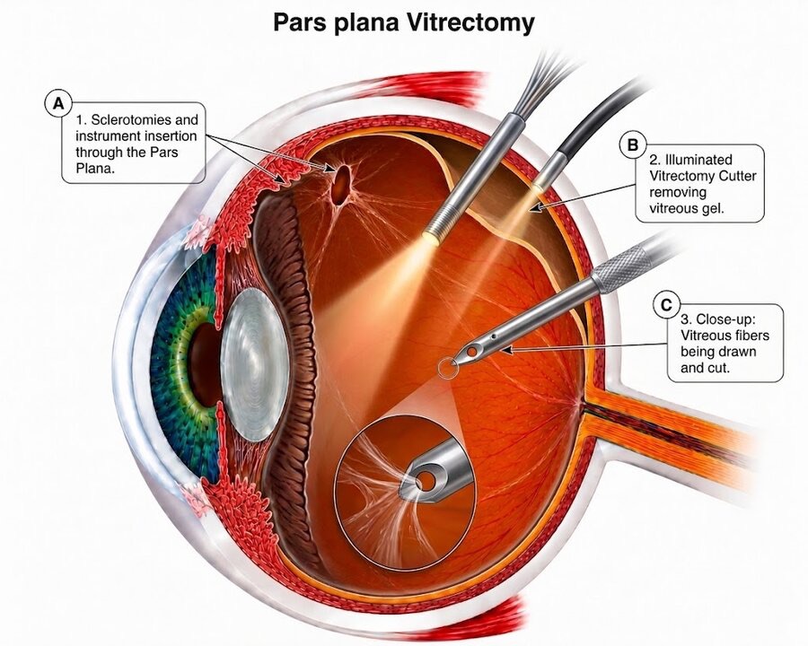
After the vitreous is removed and the retina reattached, the surgeon fills the eye with something that keeps the retina pressed in place while it heals. Two main options are used.
After vitrectomy with gas or oil, your surgeon will ask you to maintain a face-down (prone) position for several hours a day. This sounds difficult — and it is — but it is one of the most important things you can do for the success of your surgery.
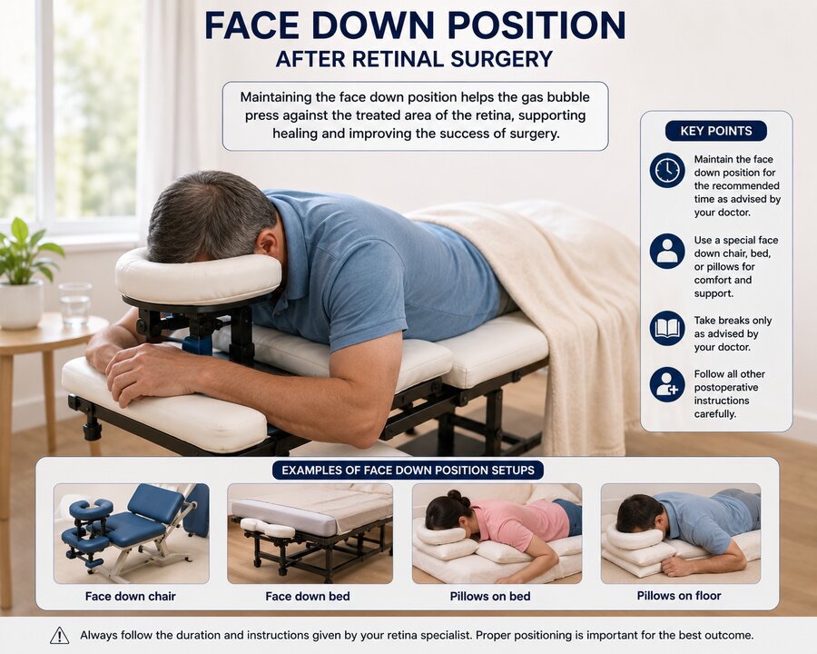
Gas and oil are both lighter than the fluid in the eye, so they float upward. After surgery, the tear is usually at the upper back of the eye. When you face down, the bubble rises and presses directly against the treated area, holding the retina snugly in place while the laser seal forms. Without this positioning, the bubble floats away from the repair and the retina may not heal properly.
A padded face rest you sit in like a massage chair — watch TV or listen to music.
A padded table with a hole for your face — like a massage table — resting without neck strain.
Pillows arranged to support your face and chest while lying down — a good affordable option.
For those who prefer the floor — pillows arranged for a comfortable face-down rest.
We know this is the question closest to your heart: "Will I be able to see normally again?" The honest answer needs a little nuance — and you deserve the truth, told kindly.
If the macula was still attached at surgery, recovery is usually much better. If it had already come off, central vision may remain limited even after successful surgery.
Like a plant pulled from soil — the longer it's disconnected from its blood supply, the more the cells suffer. Time matters enormously.
A small, early detachment heals better than an extensive one involving most of the retina.
Pre-existing conditions like diabetes, severe near-sightedness, or previous eye surgery affect the final outcome.
Yes — in a minority of cases, the retina can detach again even after successful surgery. This is not a failure of the surgery; it is a known risk your doctor will discuss with you.
Sometimes the eye forms scar tissue on or under the retina. This contracts and can pull the retina off again — called Proliferative Vitreoretinopathy (PVR), an overactive healing response that works against you.
Occasionally a new tear forms in a different area after surgery — especially in patients with widespread lattice degeneration or those who are very myopic.
There is a lot of misinformation about retinal detachment. Let's clear up the most common myths we hear from patients. Tap each one.
Old, longstanding floaters that have not changed are usually harmless. However, a sudden increase in floaters, new floaters, or floaters with flashes of light is a different situation and needs urgent examination.
Not always, but the risk can be significantly reduced. If you have high short-sightedness, a family history, or a detachment in the other eye, regular dilated check-ups are essential. Retinal tears and weak areas (lattice degeneration) found early can be sealed with preventive laser before they progress. This laser is done in the clinic, takes a few minutes, and is highly effective. Patients with high myopia, those who have had cataract surgery, and those with a strong family history should specifically ask whether preventive laser is advisable — a timely session can often prevent the need for surgery altogether.
Most retinal surgeries are done under local anaesthesia (only the eye is numbed) with sedation so you are relaxed and comfortable. General anaesthesia may be used in children or specific cases. Your surgeon and anaesthetist decide what is best for you.
Most surgeries are done as a day-care procedure — you come in the morning and go home the same evening. In complex cases, an overnight stay may be advised.
Light activities can usually resume within a week or two. Strenuous activities, heavy lifting, swimming and rubbing the eye should be avoided for at least 4–6 weeks, or as advised. Reading and watching TV can usually resume within days.
Your glasses prescription may change after surgery, especially after vitrectomy. Wait until the eye has fully settled (usually 2–3 months) before getting new glasses prescribed.
Yes. You can use a face-down chair or table and watch TV placed below you on the floor. Phones can be used while facing down. Many patients find audiobooks, podcasts and music helpful during this period.
Yes, there is no restriction on using your other eye. However, it is important to have it checked carefully too, as it has a higher risk of developing tears or detachment.
Yes, absolutely. Children and young adults with high myopia should have regular dilated retinal examinations. Early detection of weak areas, lattice degeneration or retinal holes allows preventive laser before any damage occurs.
Oil removal is a relatively shorter, safer procedure compared to the original detachment surgery. It is usually done about 2–4 months after the primary surgery, once the retina is confirmed stable and well-attached.
Signs of success include gradual improvement in vision, absence of the dark curtain or shadow, and a normal-looking retina on examination. Your surgeon monitors this at each follow-up visit.
It is completely natural to feel anxious. Please talk to your surgeon and care team — we are here to answer every question and every fear. This surgery is designed to save your vision, and modern retinal surgery has excellent outcomes. Your eye is in good hands.
Most mediclaim and health insurance policies do cover retinal detachment surgery — it is a medical emergency, not a cosmetic procedure. However, the exact coverage, cashless facility and claim process vary from one policy to another. We strongly recommend that you or your family confirm the details directly with your insurance company before or at the time of admission. Our hospital staff will be happy to help with the documents and paperwork to support your claim. Read our simple Insurance & Cashless Guide →
Our retina clinic, surgeries & specialists at Bapaye Eye Hospital
How cashless works, coverage, co-payment & claims — simply explained
Check if your insurer or TPA is on our cashless panel
See a retina specialist — walk-in 10am–8pm, or book online
Walk-in: 10 am – 8 pm · Call 9423971283 · Emergency 24×7
Book an Appointment